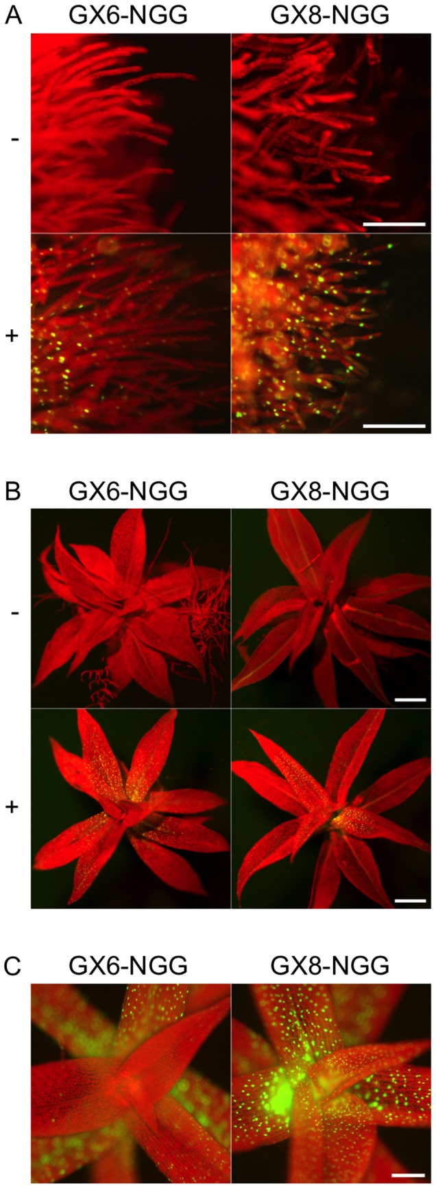
Figure 2. Spatial expression patterns of NGG induced by P . patens XVE system.

Fluorescence images of protonemata (A) and gametophores (B) of GX6-NGG#63 and GX8-NGG#4 lines. Fluorescent signals were observed after 24 h inoculation in water with (+) or without (-) 1 µM β-estradiol. Yellowish color of leaf veins in GX8-NGG#4 is caused by reflection but not by NGG signals. (C) Magnified views of apexes of gametophores in GX6-NGG#63 and GX8-NGG#4 lines. Pictures in A, B, and C panels are at the same magnification. Bars = 100 µm.
