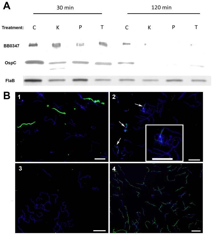
Figure 4. BB0347 is surface exposed in B. burgdorferi MI-16.
A) Intact spirochetes were treated with different proteases for 30 min or 2 hrs. Western blots were run against whole cell lysates after these treatments and blotted with αBB0347, αOspC, or αFlaB. After two hours, no difference was observed between the control and protease-treated spirochetes in the αFlaB-blotted membranes, but OspC and BB0347 were almost completely degraded (Key: C: No protease control, K: proteinase K, P: pronase, T: trypsin). B) Intact bacteria were coated onto glass slides and fixed with 10% Formalin. Antibodies against the same proteins listed in (A) were used to stain and a secondary antibody conjugated with Dylight 488 was used for detection. DAPI was used as a secondary stain to localize spirochetes (see Materials and Methods). An additional control in which the B. burgdorferi membrane was compromised by desiccation, was included to verify that αFlaB antibodies were effective on fixed spirochetes. Key: Panel 1) αOspC, 2) αBB0347, 3) αFlaB 4) αFlaB with membrane disruption. BB0347 was detected in intact spirochetes, further verifying the surface exposure. White bars indicate a length of 10 µM, and the magnification is 1000×. Results are indicative of three independent experiments with similar outcomes.

