
Figure 2. Localisation of infarction.
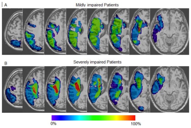
Overlap of infarct lesions on a T1 anatomical template in (A) mildly impaired patients (n=14) and (B) severely impaired patients (n=8). Colour bar indicates the proportion of patients with infarction for each voxel.