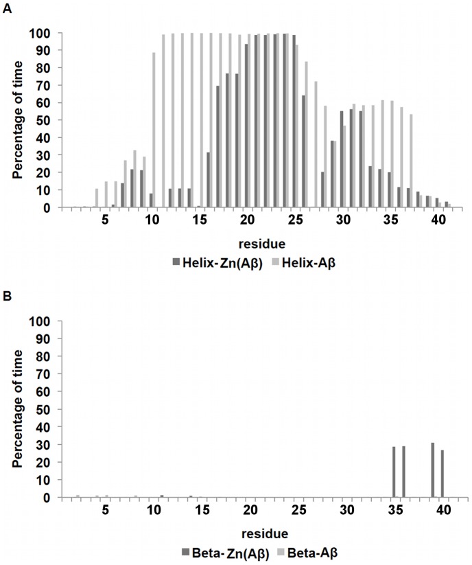
Figure 7. Per-Residue Secondary Structure of Zn(Aβ) and Control.
The percentage of time (after equilibration) peptide residues adopt helix or β-sheet structure is shown. (A) shows the per-residue helix. (B) shows the per-residue β-sheet. Dark gray lines are Zn(Aβ) where Zn binds at Glu11 His6, 13 14. Gray lines are the control. The peptide has the same initial structure in the Zn(Aβ) complex and the control simulation.

