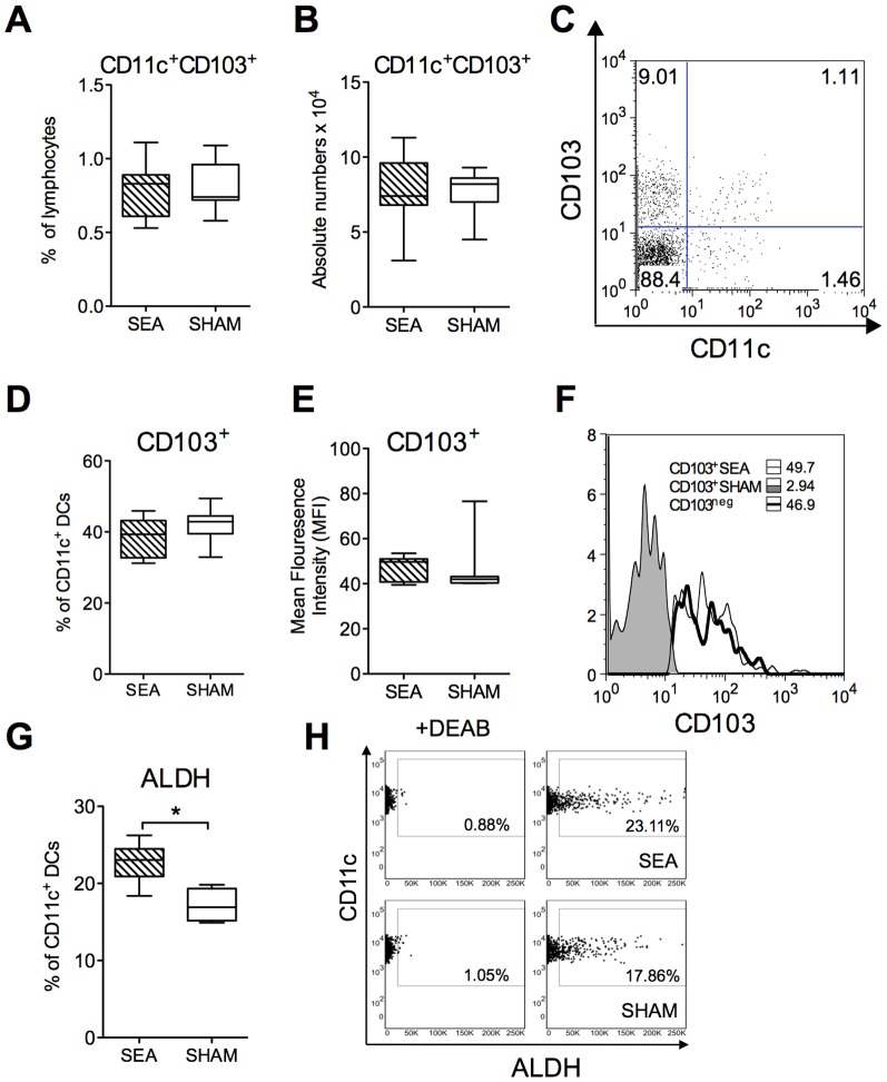
Figure 3. Increased expression of ALDH by CD11c+ DCs after neonatal SEA treatment.
Mice (n = 6 in each group) were fed staphylococcal enterotoxin A (SEA) or PBS (SHAM) perorally on six occasions during the first two weeks of life. Mesenteric lymph nodes (MLNs) were collected at four weeks after treatment and the cells were subjected to flowcytometric analysis. A) The proportion of CD11c+CD103+ DCs among live-gated MLN cells. B) The absolute number of CD11c+CD103+in MLN. C) gating of CD11c+ CD103+ DCs in MLN. D) Proportion of CD103+ cells among the CD11c+ DC population. E) Mean Fluorescence Intensity (MFI) of CD103 on the CD11c+ DC population. F) Representative histogram of CD103 expression on CD11c+DC population. The ALDEFLUOR® assay was performed in order to identify CD11c+ cells with aldehyde dehydrogenase (ALDH) expression (ALDH+). The ALDH inhibitor diethylaminobenzaldehyde (DEAB) was used as a negative control. The cells without inhibitor, shifted to the right, were considered ALDH+ cells. (G) The proportion of CD11c+ cells expressing ALDH. (H) Representative dot plot of MLN cells stained with CD11c and ALDH (right panel) and with DEAB (left panel). Error bars represent SD and horizontal line shows the median value of the group. Data are representative from two independent experiments. * P<0.05, analyzed with Mann-Whitney U-test.

