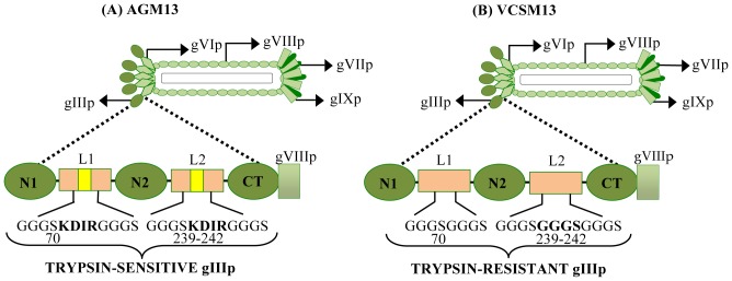
Figure 1. Schematic representation of gIIIP protein encoded by the helper phages AGM13 and VCSM13.

The N1, N2 and CT domains of the coat protein gIIIP are depicted as oval boxes (green) and are joined by the linkers L1 and L2. (A) A four-amino acid ‘KDIR’ trypsin cleavage site was introduced into both L1 and L2 linkers in the AGM13 helper phage, to encode trypsin-sensitive gIIIP. (B) VCSM13 is the wild-type helper phage without any trypsin cleavage sites, which encodes a trypsin-resistant gIIIP. The ‘KDIR’ amino acid sequence was inserted after residue 70 of gIIIP in L1 (shown in bold in A) and replaced four amino acids between residues 239–242 of gIIIP in L2 (shown in bold in B).
