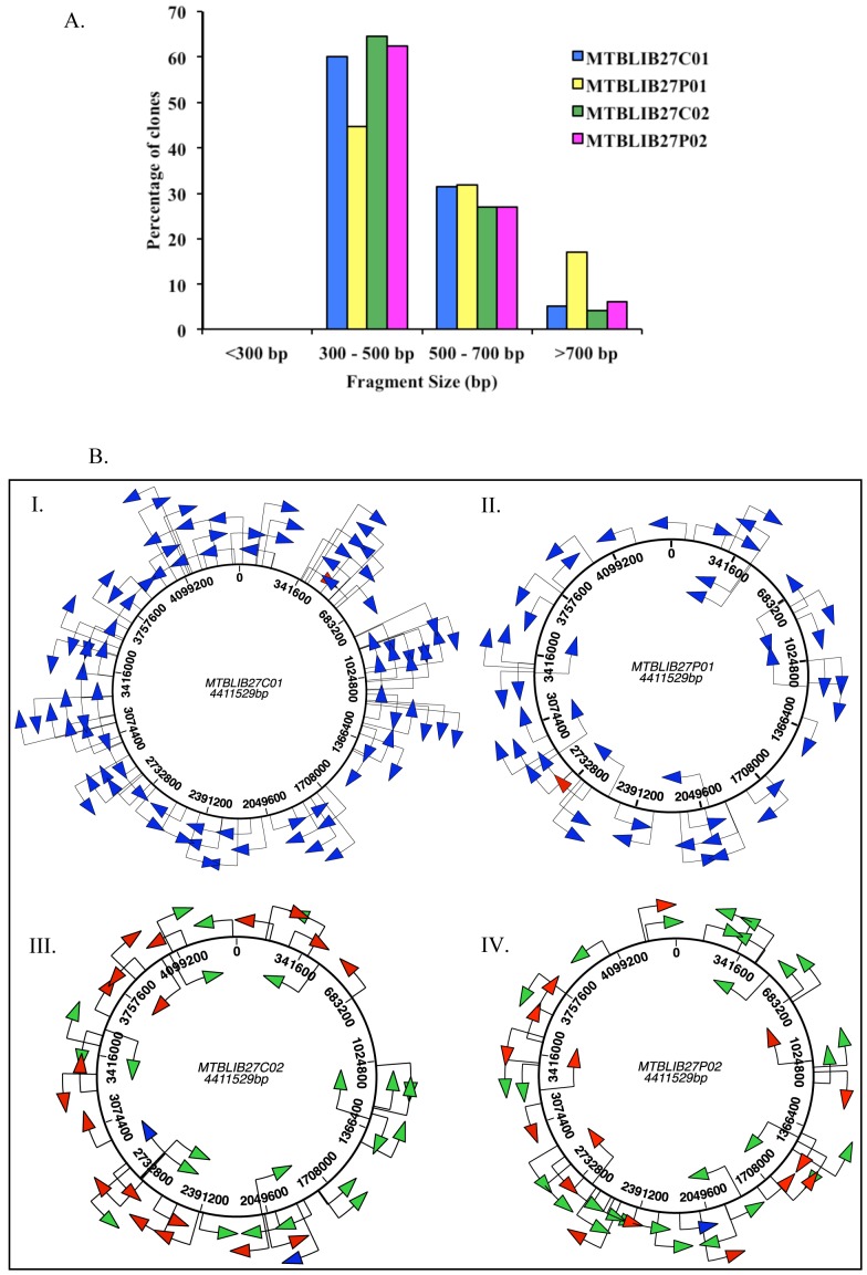
Figure 5. Distribution of M. tuberculosis H37Rv gene fragments in the MTBLIB27 library.
5A. Bar graph depicting the size distribution of M. tuberculosis H37Rv gene fragments in the MTBLIB27 at different stages of library construction. 5B. Schematic representation of the distribution of gene fragments. The M. tuberculosis genome is ∼4.4 Mb and consists of ∼4000 genes. (I) MTBLIB27C01 primary cells before ORF selection; (II) MTBLIB27P01 after trypsin treatment (10 µg/ml) of primary phages; (III) MTBLIB27C02 secondary cells obtained after trypsin treatment (10 µg/ml) of primary phages for ORF selection; (IV) MTBLIB27P02 obtained from rescue of C02. The non-ORF selected inserts which align with the M. tuberculosis genome are shown as blue arrows; the clones in-frame with PelBss and gIIIP are indicated as red arrows (non-genic clones); the clones aligning with the M. tuberculosis proteome are indicated as green arrows (genic clones). The direction of the arrows indicates gene orientation. The maps are to scale.

