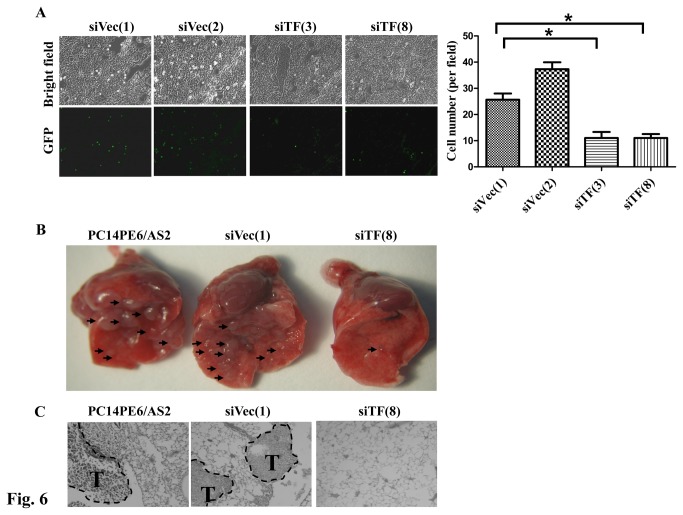
Figure 6. Blockage of TF expression decreased cell adhesion and lung metastasis in nude mice.
(A) PC14PE6/AS2-siTF (siTF(3) and siTF(8) and PC14PE6/AS2-siVec cells (siVec(1) and siVec(2)) stably expressing Green fluorescent protein (GFP) were applied to the frozen lung sections. The glass slides were shocked at 70 rpm for 20 min. After PBS wash, adhering cells were fixed and photographed by fluorescent microscopy (left panel). The cell number was quantified as right panel. (B) Various cells (1 × 10⁶) such as parental (PC14PE6/AS2), vector control (siVec(1)), or siTF-transfected PC14PE6/AS2 cells (siTF(8)) were suspended in 0.1 ml PBS and then injected intravenously into the tail veins of nude mice. Mice were sacrificed and lungs were excised and photographed 26 days after injection. Block arrows: metastatic tumor nodules. (C) Histological analysis of lung metastasis of PC14PE6/AS2, siVec(1) or siTF(8) cells. Paraffin-embedded lung tissues were sectioned into 4 µm thick sections, and then stained with hematoxylin-eosin. Metastatic tumors (T) are shown within the PC14PE6/AS2 and siVec(1) lung tissue. The siTF(8) tumor had no foci.

