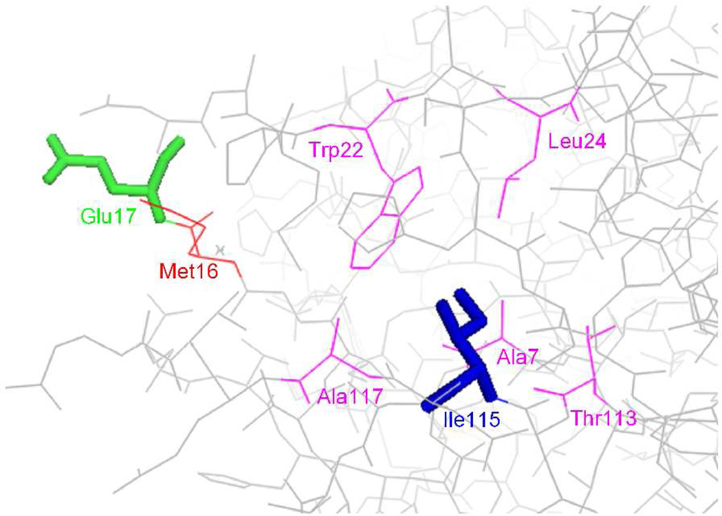
Figure 1.
Structure of wild-type E. coli DHFR structure (PBD 1RA1), showing Glu17 in green and Ile115 in blue. The amino acid residue (Met16) which is close spatially to the side chain of Glu17 (≤ 4 Å) is shown in red. The amino acid residues (Ala7, Trp22, Leu24, Thr113 and Ala117) which are close to the side chain of Ile115 (≤ 4 Å) are shown in magenta.

