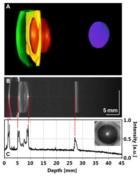
Figure 2.
Full eye length imaging using swept source optical coherence tomography. A, 3-D rendering of the volumetric data set (cornea – green, iris – yellow, crystalline lens – orange, retina – blue). B, Central cross-section. C, Extracted profile enables identification of ocular surfaces allowing for measurements of intraocular distances.

