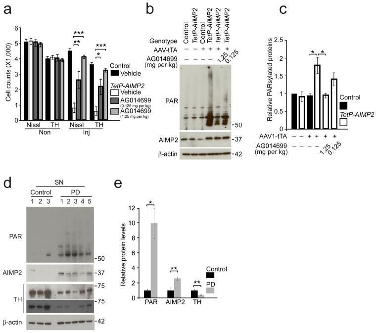
Figure 6.
PARP inhibition protects against AIMP2 induced dopaminergic toxicity.
(a) Stereological assessment of tyrosine hydroxylase positive and Nissl positive dopaminergic neurons in the SNpc of control and TetP-AIMP2 mice 25 days following AAV-tTA injection with or without treatment with the PARP inhibitor, AG014699 (Vehicle, 50% DMSO in saline; 0.125 mg/kg or 1.25 mg/kg AG-14699, bid, i.p.), −3 days through 25 days after the viral injections (n = 3 per group).
(b) AIMP2, and PAR conjugated proteins in the ventral midbrain of control and TetP-AIMP2 mice 16 days following intranigral injection of AAV-tTA with the treatment of AG014699 monitored by western blot.
(c) Quantification of PAR conjugated protein levels in panel (b) normalized to β-actin (n = 3 for 1.25 mg/kg AG-14699 treatment group, and n = 4 for the other groups).
(d) PAR conjugated proteins, and AIMP2 in the substantia nigra of postmortem brain tissues from Parkinson’s disease (PD) patients and controls monitored by western blot. Extent of dopamine neurodegeneration was determined by anti-tyrosine hydroxylase immunoreactivity.
(e) Quantification of PAR conjugated protein levels in panel (d) normalized to β-actin.
Quantified data (a, c, e) are expressed as mean ± s.e.m., *P < 0.05, **P < 0.01, ***P < 0.001, ANOVA test followed by Student-Newman-Keuls post-hoc analysis (a), Kruskal-Wallis ANOVA test (c), unpaired two-tailed Student’s t-test (e). Full length blots are presented in Supplementary Figure 14 (b, d).

