Figure 2.

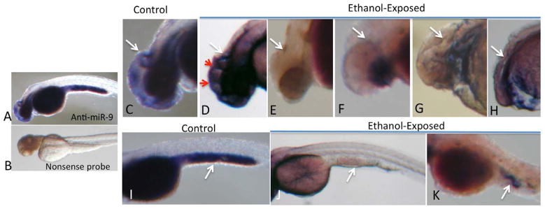
In situ hybridization for miR-9 at 48 hpf. Specific hybridization, visualized by alkaline-phosphates linked histochemistry (blue color product), was detected with an anti-miR-9 probe (A) but not with a nonsense control probe (B). In control zebrafish, specific hybridization was localized to the brain (D) and yolk sac (I). Within the brain (C), hybridization was observed in telencephalon and diencephalon, with a prominent hybridization ridge (white arrow) adjacent to the midbrain/hindbrain boundary. Ethanol exposure resulted aberrant miR-9 expression along ectopic ridges (D, red arrows) in zebrafish exhibiting the least dysmorphology. Ethanol exposure also resulted in a range of microcephaly, microphthalmia and anophthalmia (D–H), with a complete loss in neural miR-9 expression, including loss of hybridization adjacent to the midbrain/hindbrain boundary in microcephalic zebrafish. MiR-9 expression was also observed in yolk sac (I, white arrow points to hybridization in the posterior yolk sac). Ethanol exposure results in loss of miR-9 expression (J,K).
