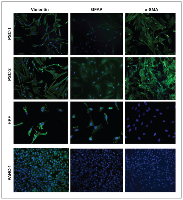
Figure 2.
Pancreatic stellate cells are positive for GFAP and α-SMA. Immunofluorescence analysis of representative primary PSC lines grown on chamber slides. Cells were stained with 4′, 6-diamidino-2-phenylindole (DAPI; blue) and antibodies (green) for vimentin, GFAP, and α-SMA. The human PANC-1 cell and HPF line were negative controls for α-SMA staining. Slides were visualized at ×40 magnification.

