Fig. 3.
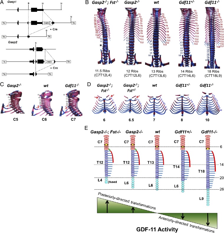
Role of Gasp2 in axial skeletal patterning. (A) Gene targeting strategy for Gasp1 and Gasp2. NEO, neomycin resistance cassette; TK, thymidine kinase. (B) Vertebral columns of WT, Gasp2−/−, Gasp2−/−;Fst−/−, Gdf11+/−, and Gdf11−/− mice. Note the changes in the number of ribs. *Gasp2−/−;Fst−/− mice also had a reduced number of lumbar vertebrae from six to four, with extensive fusion of lumbar and sacral segments. (C) Cervical and anterior thoracic regions of WT, Gasp2−/−, and Gdf11−/− mice. Note the shift of the position of the anterior tuberculum (arrow) from C6 to C5 in Gasp2−/− mice and from C6 to C7 in Gdf11−/− mice. (D) Vertebrosternal ribs of WT, Gasp2−/−;Fst+/−, Gasp2−/−;Fst−/−, Gdf11+/−, and Gdf11−/− mice. Note changes in the number of attached ribs and additional cervical ribs (arrow). (E) Schematic representation of vertebral columns. Gdf11+/− and Gdf11−/− mice have anteriorly directed homeotic transformations of the axial skeleton. In contrast, mice lacking certain GDF-11 inhibitors (Gasp2−/− and Gasp2−/−;Fst−/−) have posteriorly directed transformations. Cervical (orange), thoracic (purple), and lumbar (sky blue) vertebrae, anterior tuberculi (small blue dots), ectopic ribs from a cervical vertebra (green curved lines), sternums (red curves), and ribs (blue lines) are color coded as indicated. The dashed lines indicate the normal positions of typical vertebral characteristics: 6 for the anterior tuberculum, 14 for the most posterior rib attached to the sternum, 20 for the most posterior thoracic vertebra, and 26 for the most posterior lumbar vertebra.

