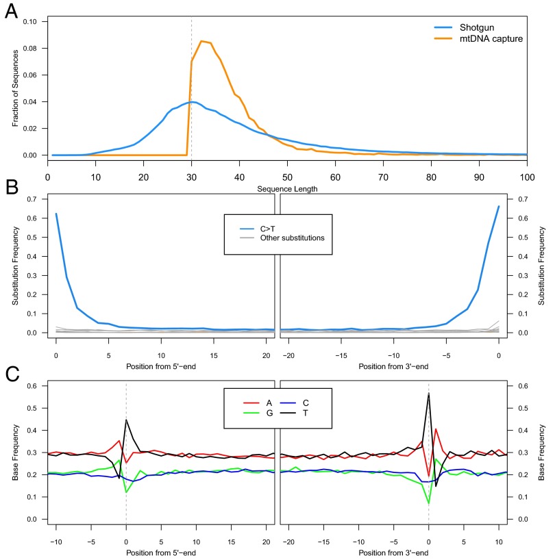
Fig. 2.
Length distributions and damage patterns. (A) Fragment length distributions of shotgun sequences (blue) and captured mitochondrial sequences (orange) as the fraction of sequences in each size bin. (B) Substitution patterns at the 5′ and 3′ ends of the aligned sequences. (C) DNA fragmentation patterns inferred from the reference base composition around alignment start and end points.

