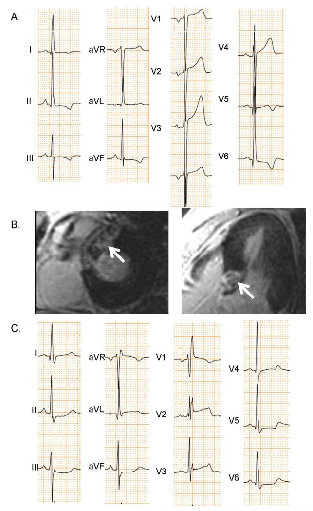
Figure 4. ECGs and CMR-LGE of Patient Developing RBBB after Alcohol Septal Ablation.
This hypertrophic cardiomyopathy patient had a baseline ECG (A) showing left ventricular hypertrophy, but not bundle branch block. Alcohol septal ablation was performed in a proximal LAD septal perforator leading to necrosis highlighted by arrows in the CMR-LGE image (B). Post septal ablation ECGs (C) showed that RBBB developed.

