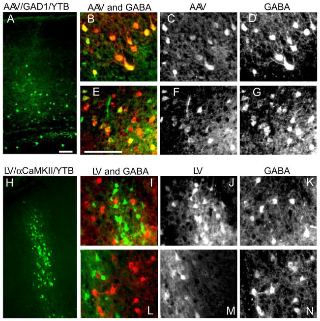
Figure 3. Cell-type Specificity of Helper-Virus Transduction in Mouse Neocortex.
Low magnification images show expression of YFP (green) in cortical neurons following AAV/GAD1/YTB (A; AAV) and LV/αCaMKII/YTB (H; LV) helper-virus injections in mouse cortex. Higher magnified black and white images of AAV infected neurons (C and F) co-localize with neurons labeled through GABA immunofluorescence (D and G), as can be seen when shown together in color (B and E; AAV is shown in green and GABA in red). In contrast, higher magnified black and white images of LV infected neurons (J and M) do not co-localize with neurons positive for GABA immunofluorescence (K and N), as can be seen when shown together in color (I and L; LV is shown in green and GABA in red). Scale bars = 100 μm. See also Fig. S1.

