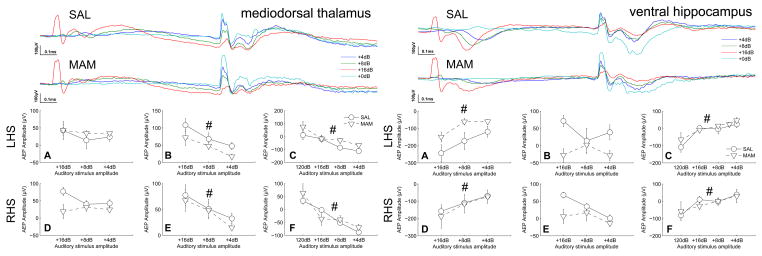
Figure 4.
No deficits in auditory information processing were seen in the mediodorsal thalamic nucleus or ventral hippocampus of MAM rats when compared with SAL treated controls. However, the mediodorsal thalamic nucleus, a major origin of cortical efferents, appeared to play a role in intensity processing at a similar latency to that seen in the prelimbic cortex. The ventral hippocampus also appeared to play a role in intensity processing. Both the mediodorsal thalamus and the ventral hippocampus display an amplitude dependent modulation of the response to the startle-stimulus. (Top traces of each subfigure) Grand averaged AEPs for SAL and MAM treated animals respectively. (A,D) Earlier occuring maximum evoked potential amplitudes and (B,E) later occuring maximum evoked potential amplitudes for the prepulse intensities. (C,F) Evoked potential amplitudes of the P20 component of the potentials evoked to the startle-stimulus during both prepulse and startle alone trials (+0dB). * indicates a significant effect of treatment between SAL and MAM animals. # indicates a significant effect of prepulse intensity. Data are plotted as the mean ± SEM.

