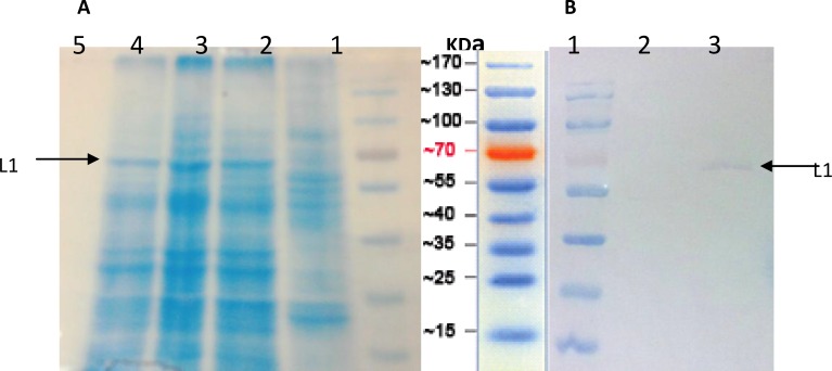
Figure 3.
SDS-PAGE and Western blot analysis of HPV16- L1 protein expressed in Sf9 cells. (A) SDS-PAGE.. Lane 1; Protein Marker, Lane 2; uninfected Sf9 cells, and Lane 3, 4 and 5; Sf9 cells transfected with recombinant HPV16-L1 gene bacmid DNA. (B) Western blot analysis using the anti-HPV16-L1 monoclonal antibody. Lane 1; Protein marker, Lane 2; uninfected Sf9 cells, and Lane 3; Sf9 cells transfected with recombinant HPV16-L1 gene bacmid DNA. As shown un-specific bands didn’t observed using monoclonal antibody

