
Fig. (1).
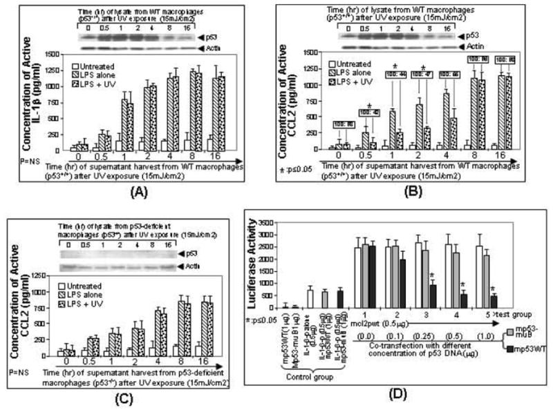
Regulation of LPS-induced CCL2 production by the accumulation of endogenous p53. Mouse primary macrophage cells from wild-type mice (A, B) or p53-deficient mice (C) were untreated as control (white bar) or treated with LPS (downward diagonal striped bar) or LPS plus UV radiation (dotted bar). Cells were then cultured. The medium from each group of treated cells were harvested at different times (0, 0.5, 1, 2, 4, 8 or 16 hrs) and assessed for IL-1-β production as control (A) or CCL2 production (B, C) by ELISA (n = 3). The concentration of CCL2 secreted from LPS-treated cells was assigned a value of 100% as the baseline, and the relative CCL2 concentration from co-treated cells by LPS plus UV was calculated (boxed ratio). Values were normalized with respect to protein concentrations. Data are presented as mean ± SEM. In order to detect p53 accumulation, the lysate from cells at each time point was purified and analyzed by Western blot with the antibody against p53 or actin as control (Top panel of A-C). D. Regulation of CCL2 promoter activity by overexpression of p53. U2OS cells were transfected with reporter DNA (mcl2pwt alone or mouse IL-1β promoter alone, white bar, control group) or co-transfected with mcl2pwt DNA and different concentrations (0, 0.1, 0.25, 0.5, or 1μg) of p53 DNA (grey bar for mp53WT or black bar for mp53-muB as control, experimental group 1-5). Cells were cultured overnight. The lysate from cells at each group was assessed by luciferase assay (n = 3). Data are presented as mean ± SEM.