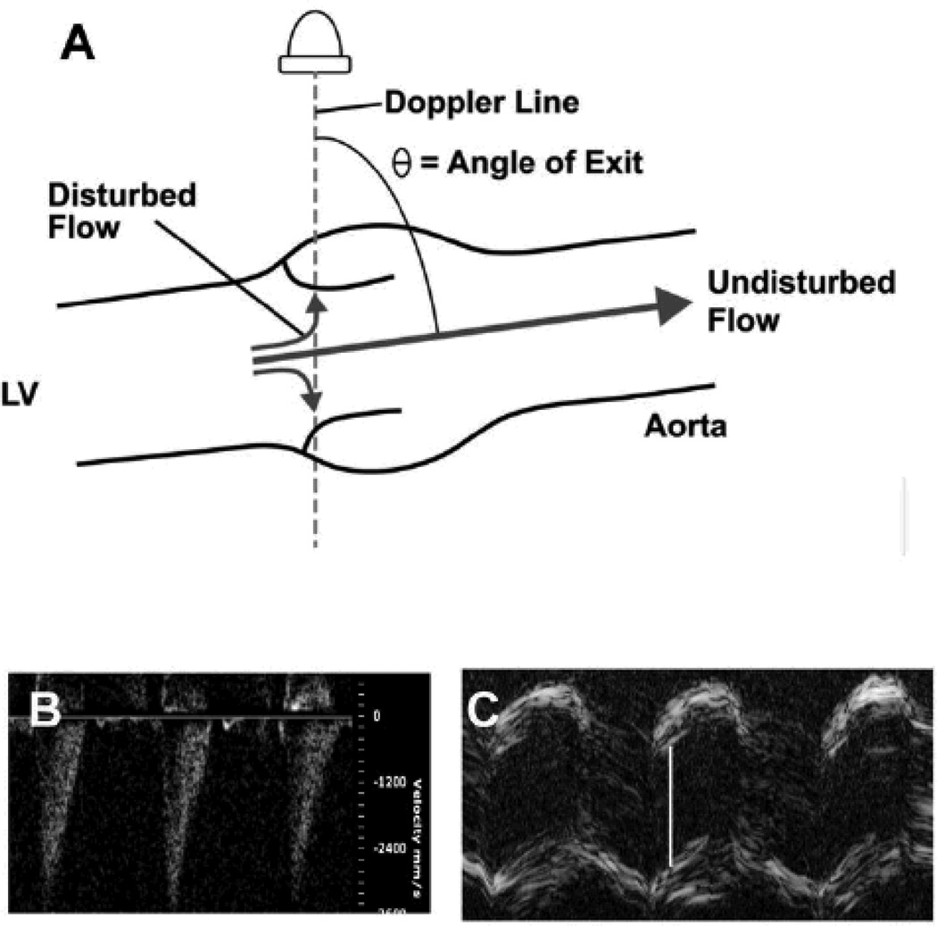
Figure 2. Overestimation of stenosis severity.
A. Calculation of transvalvular blood flow velocity requires angle correction when the direction of flow is not colinear with the line of Doppler interrogation. In the illustration, the angle of exit of presumed blood flow is 75° . Thus, measured velocity would be divided by the cosine of 75°, = 0.26, in order to compute actual flow velocity. When transvalvular blood flow is disturbed, a portion of the flow jet will be directed more parallel to the Doppler line, and will be detected as a higher velocity. In this case, correction for a presumed angle of exit of 75° will overestimate true blood velocity by a factor of about 4. B. Doppler velocimetry from a normal mouse, with neither aortic stenosis nor regurgitation. The angle between the Doppler cursor and the presumed direction of transvalvular flow was 75° (image not shown). Peak velocity is reported as = 3.6 m/s, erroneously predicting a peak valve gradient of 52 mmHg. C. M-mode echocardiogram of the aortic valve in the same mouse. White bar indicates aortic cusp separation = 1.3 mm, which is normal. Color Doppler and magnetic resonance imaging both confirmed the absence of aortic regurgitation (images not shown).

