
Figure 4.
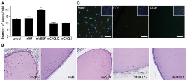
Comparison of the tube formation potential of the angiogenic factors/chemokines in vivo. a In vivo tube formation in transplanted Matrigel plugs containing MIF, VEGF, CXCL12, or CXCL1 analyzed after 1 week (*p < 0.05 vs. control; n = 5). b Representative H&E stains from a (scale bar 400 μm). c Specific immunohistochemistry stainings for macrophages (Mac-2), lymphocytes (CD3) and endothelial cells (CD31) as detected in MIF-containing Matrigel plugs (scale bar 25 μm)