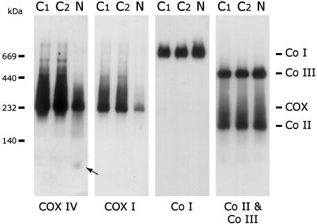
Figure 2.
Blue-Native PAGE analysis of COX levels in patient heart mitochondria. Heart mitochondria (10 μg protein) from two control subjects (C1 and C2) and the patient (N) were separated by BN-PAGE, and the content of COX, Complex I (Co I), Complex II (Co II), and Complex III (Co III) were determined by immunoblot analysis using antibodies against COX subunits I and IV (COX I and COX IV), the 39-kDa subunit of complex I, the 70-kDa subunit of complex II, and the Core1 protein of complex III. Patient heart mitochondria show a specific reduction in the total amount of fully assembled COX but no evidence of any subcomplexes. The arrow indicates the small amount of unassembled COX IV in the patient. The migration of molecular mass standards is indicated on the left.

