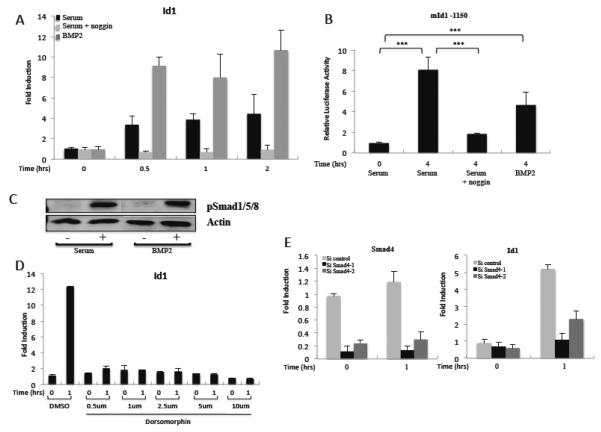
Figure 6. The BMP pathway is necessary and sufficient for serum induction of Id1.
A) NIH3T3 cells were serum-starved overnight and induced with 20% serum or 20 ng/ml BMP2 as indicated. Where indicated, 100 ng/ml noggin was added during serum-starvation. Gene expression was measured by qPCR as in figure 1. B) NIH3T3 cells were transfected with the mId1 −1150 and pRLSV40P luciferase reporter constructs, serum starved with or without noggin, and induced with serum or BMP2 as in A. Luciferase levels were measured as in figure 3. C) NIH3T3 cells were starved in 0.2% serum overnight then induced with 20% serum or 20 ng/ml BMP2 for 1 hour. The lysates were immunoblotted with phospho-Smad1/5/8 specific antibodies or anti-actin as a loading control. D) NIH3T3 cells were serum-starved, treated with Dorsomorphin at the indicated concentrations for 1 hour, and then induced with 20% NCS for 1 hour. Id1 mRNA levels were measured by qPCR as in figure 1. E) Cells were transfected with control or Smad4 siRNAs. The next day the media was changed to 0.2% serum overnight and the cells serum-induced for 1 hour. Smad4 and Id1 mRNA levels were measured by qPCR as in figure 1.

