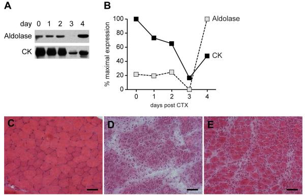
Figure 3. Aldolase levels are high in early regenerating muscle cells in an in vivo mouse model of muscle damage and repair.
Uninjured (“day 0”) and CTX-injected anterior tibialis muscle samples were obtained by sacrificing the mice at days 1, 2, 3 and 4 post-injection/injury. (A) Lysates were made from the muscle biopsies, and equal protein amounts were immunoblotted with antibodies against aldolase A and CK. Similar data were obtained in 2 separate experiments. (B) The immunoblots were quantified by densitometry as described in Figure 1 legend. (C-E) Frozen sections of uninjured mouse muscle (panel C), or muscle harvested 3 (panel D) or 4 (panel E) days after CTX injection were stained with H & E and visualized with light microscopy. Scale bar: 50 microns. CK: creatine kinase; CTX: cardiotoxin.

