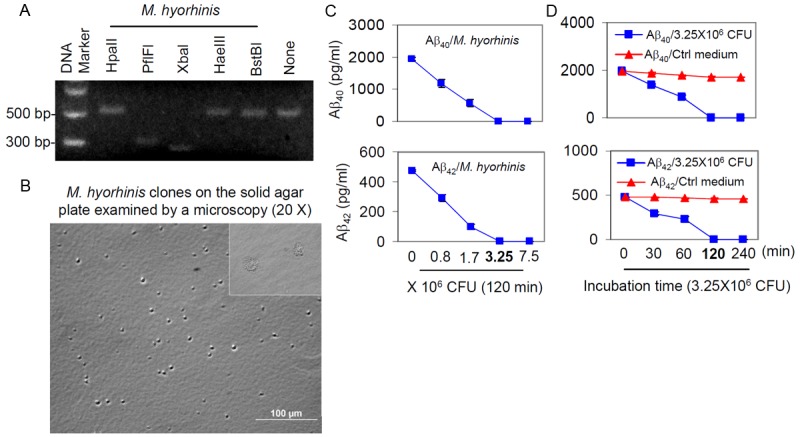
Figure 2.

Identification of M. hyorhinis and characterization of Aβ40, 42 degradation. A: Mycoplasma genomic DNA from infected N2a cell cultures was extracted, amplified and analyzed by restriction fragment length polymorphism (RFLP) using five different restriction endonucleases. The digestion pattern observed identified the Mycoplasma as M. hyorhinis. B: M. hyorhinis colonies were observed on solid agar plate by light microscopy (20X). C: CHO/APPwt media was incubated with 0, 0.8, 1.7, 3.25, or 7.5 × 10⁶ colony forming units (CFU) of M. hyorhinis at 37°C for 120 min. D: CHO/APPwt media was incubated with 3.25 × 10⁶ CFU of M. hyorhinis or clean Mycoplasma media (Ctrl Medium) for 0, 30, 60, 120 and 240 min at 37°C. Aβ40 and Aβ42 were then determined in the media by ELISA and presented as mean ± SD. These results are representative of three independent experiments with three replicates for each condition.
