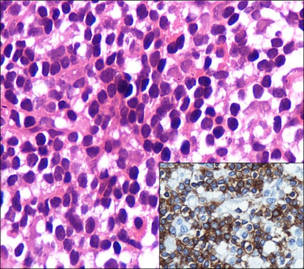
Fig. 2.

Photomicrograph showing hypercellular marrow with diffuse infiltration by lymphoid cells, plasmacytoid lymphocytes, a few plasma cells, and mast cells (hematoxylin and eosin stain, ×1,000); inset photomicrograph showing strong cytoplasmic positivity for CD20 in the majority of the lymphoid cells (immunohistochemical stain for CD20, ×400).
