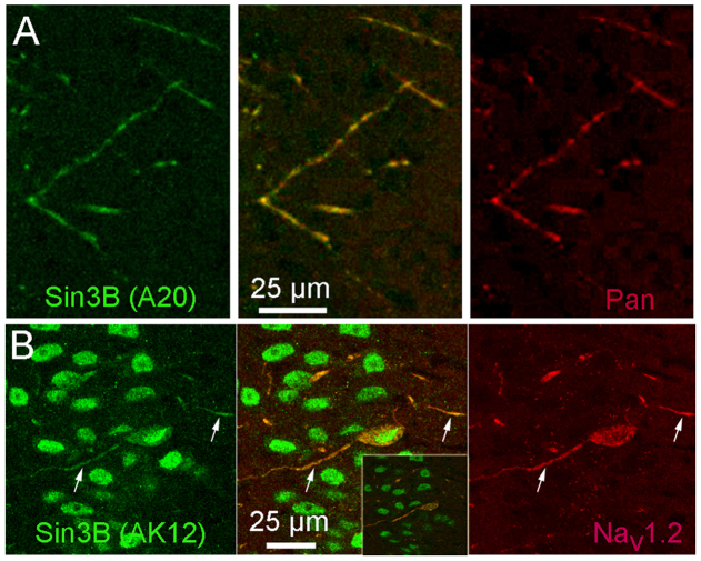
Figure 4. Sin3B immunoreactivity is found outside the nucleus, where it colocalizes with immunostaining for Nav channels A.
Discrete processes in the molecular layer of region CA1 of the hippocampus were labeled with anti-Sin3B (A20, green) as well as with a pan-specific antibody against Nav channels (Pan, red). The middle panel shows superposition of the anti-Nav-channel and anti-Sin3B staining. Images are single confocal optical sections. B. Extranuclear staining with anti-Sin3B (AK12, green) colocalized with immunostaining for Nav1.2 (red). Arrows indicate neuronal processes where the two immunoreactivities coincide. Images are Z-axis projections of a series of 5 consecutive confocal optical sections taken in CA1; a single optical section is shown in the inset, for comparison. Note that immunostaining for Sin3B in panels A and B used two different anti-Sin3B antibodies.

