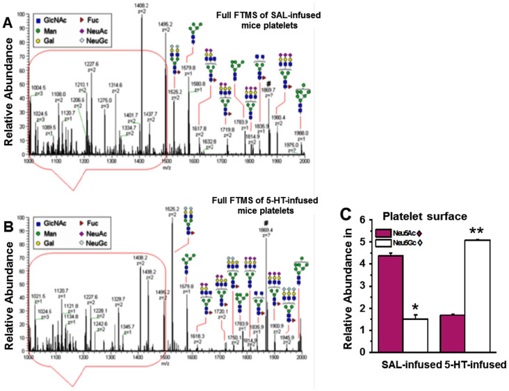
Figure 2. NSI-MS spectra of permethylated N-glycans from platelets of saline (SAL) (A) and 5-HT-infused (B) mice.
An equal number of platelets from WT mice infused for 24 hr with SAL or 5-HT were collected and plasma membrane (PM) was isolated34. The N-linked glycans on membrane vesicles were released enzymatically by PNGase F. Released N-glycans were permethylated and profiled by an LTQ Orbitrap Discoverer mass spectrometer equipped with a nanospray ion source. Glycans are detected as singly [M + Na]+, doubly [M + Na]2+ and triply [M + Na]3+ charged species. Structural assignments are based on MS/MS fragmentation and known biosynthetic limitation38,39. (C) The relative abundance of Neu5Ac on the platelet PM of SAL-infused mice was 2.9-fold higher than Neu5Gc containing glycans, whereas the ratio for platelet PM of 5-HT-infused mice was 3-fold higher in Neu5Gc-containing glycans than Neu5Ac. The total Neu5Ac and Neu5Gc-containing glycans was not different between platelet PM of SAL and 5-HT mice.

