
Figure 4. Plasma membrane Neu5Gc detected by flow cytometry.
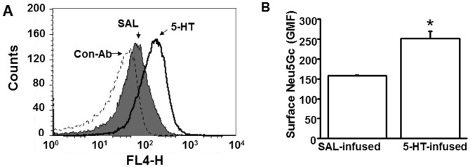
(A) The impact of plasma 5-HT on the abundance of Neu5Gc containing glycans on the platelet surface was evaluated by measuring the binding of Neu5Gc to a specific Ab. Platelets (50,000/μl) from saline (SAL) and 5-HT –infused WT mice were stained with chicken anti-Neu5Gc IgY and anti-chicken IgY DyLight 650 as primary and secondary Ab, respectively; chicken IgY was used as a control Ab. Mean fluorescence intensity of Neu5Gc expression in platelets isolated from 5-HT infused mice (black solid histogram) was higher than in platelets from SAL-infused mice (grey shaded area), black dashed histogram represents control IgY. (B) Geometric Mean of Fluorescence (GMF). Flow cytometry revealed an elevation of 33.5% in the expression levels of Neu5Gc in platelets of 5-HT-infused mice. * = statistical difference between SAL and 5-HT-infused mice.