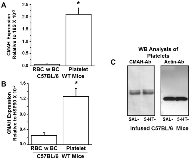
Figure 5. Expression of CMAH genes and proteins in fractionated blood samples of WT mice.
The mRNA expression levels of CMAH genes in red blood cells (RBC) and buffy coat (BC containing white blood cells) and in platelets of WT mice were normalized to the housekeeping genes 18S (A) and Hsp90 (B). The expression level of the CMAH transcript was most abundant in platelets. Primer sequences used in qRT-PCR are listed in Table 3. (C) WB analysis of CMAH in platelets revealed that CMAH protein was similarly expressed in platelets from saline (SAL) and 5-HT –infused mice. Actin was used as a loading control. Both gels were run under the same conditions.

