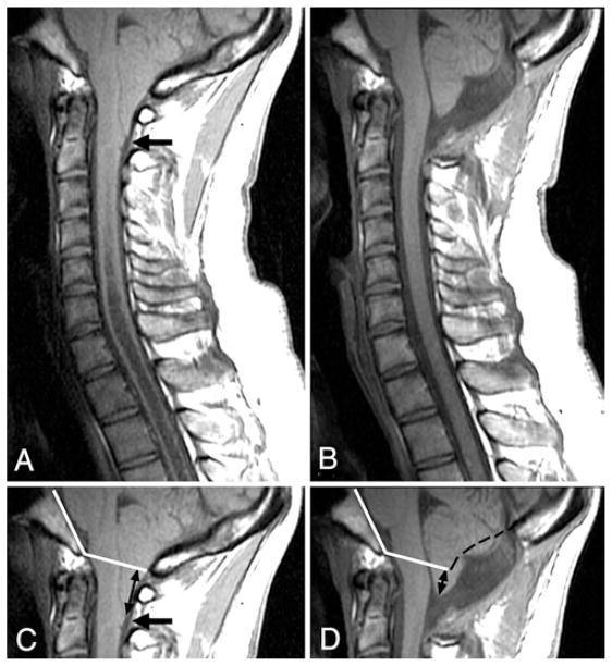
Fig. 2.
A and B Midsagittal T1-weighted MR images of the posterior fossa and cervical spine obtained before (A) and 6 months after (B) craniocervical decompression. Abnormally shaped (pointed) tonsils, dorsal cervicomedullary protuberance (arrow), and obliterated CSF spaces at the foramen magnum are evident preoperatively (A). Following surgery (B), the tonsils have assumed a normal shape, the cervicomedullary protuberance has disappeared, and CSF spaces including the foramen of Magendie have enlarged. C and D: The upper portions of the images in A and B, respectively, showing the Boogaard angle (inner angle between intersecting white lines in C and D) that was established before surgery (C) and was used to determine the level of the foramen magnum after surgery (D). In D, black dashes were added to indicate the position of the inner table of the supraocciput before surgery. Tonsillar ectopia was much reduced after surgery (D, black arrows).

