Figure 3.
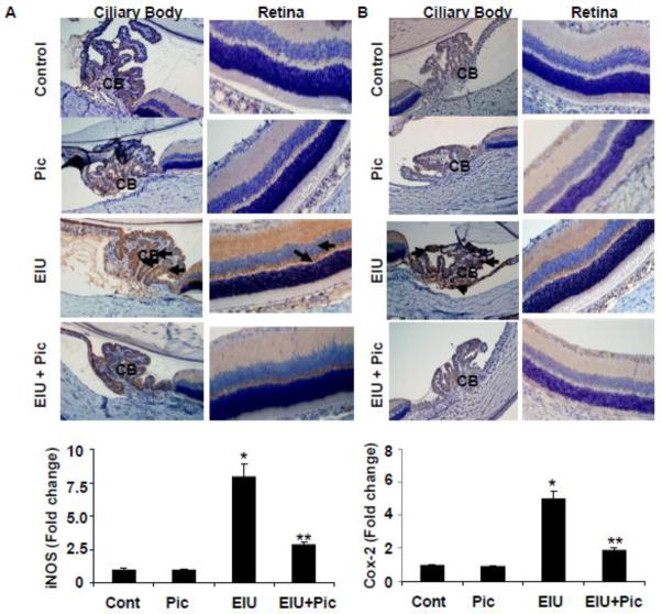
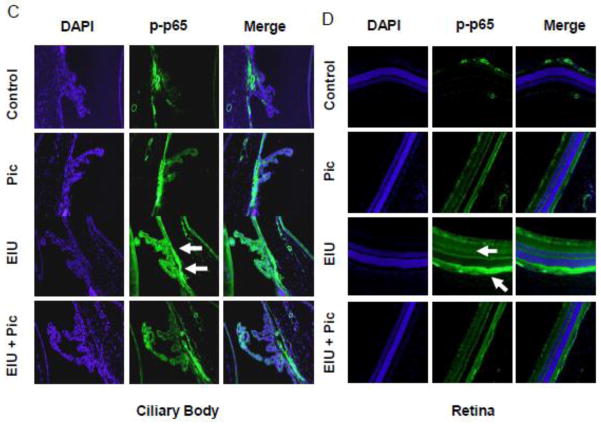
Piceatannol post-treatment prevents the expression of Cox-2 and iNOS and the activation of NF-κB in EIU. Serial sections of paraformaldehyde-fixed EIU rat eyes (24 h) were immunostained with antibodies against Cox-2 (A) and iNOS (B) and were observed under a light microscope. The bar graph shows the mRNA levels of iNOS and Cox-2 in whole rat eyes determined by RT-PCR. Data mean ± SD, n=5. *P<0.001 vs controls and **P<0.001 vs LPS. Serial sections of EIU rat eyes (3 h) were immunostained with antibodies against phospho-p65 (Ser-536)(C) followed by FITC-labeled secondary antibodies and mounted with fluorescence mounting medium with DAPI. Antibody staining intensity was observed under a Nikon fluorescence microscope for FITC and DAPI. A representative microphotograph is shown (n = 4). Arrows indicate increased expression of Cox-2 (A) and iNOS (B) as well as phosphorylation of p65 (C) in the ocular cells of ciliary bodies and retinal tissues of EIU groups. Magnification, 200x. CB - Ciliary body, R –Retina, C – Choroid, Pic – Piceatannol, EIU - Endotoxin-induced uveitis, EIU + Pic - Endotoxin-induced uveitis + Piceatannol.


