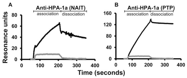
Fig. 2.

Typical SPR sensorgrams obtained with HPA-1a antibodies detectable by standard serology. Purified IgG diluted 1:8 in PBS was injected at a flow rate of 5 μL/min over HPA-1a/a and HPA-1b/b GPIIb/IIIa. The SPR signal obtained with HPA-1–negative GPIIb/IIIa was subtracted from that obtained with HPA-1a–positive GPIIb/IIIa to obtain net SPR values. (A) Pattern obtained with serum from the mother of an infant with NAIT. (B) Pattern obtained with serum from a patient with post-transfusion purpura. Gray tracing is signal obtained with serum from a normal individual.
