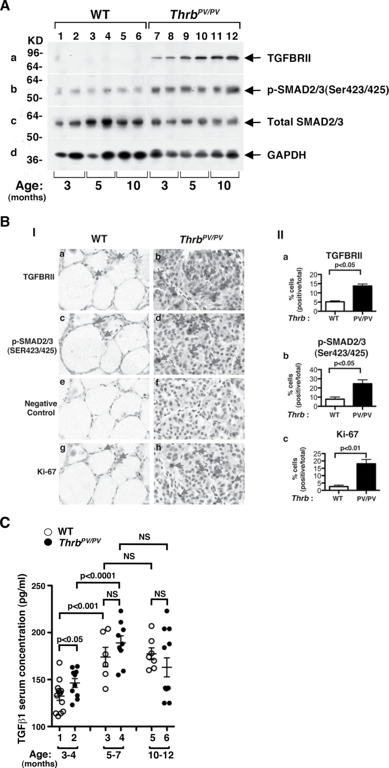
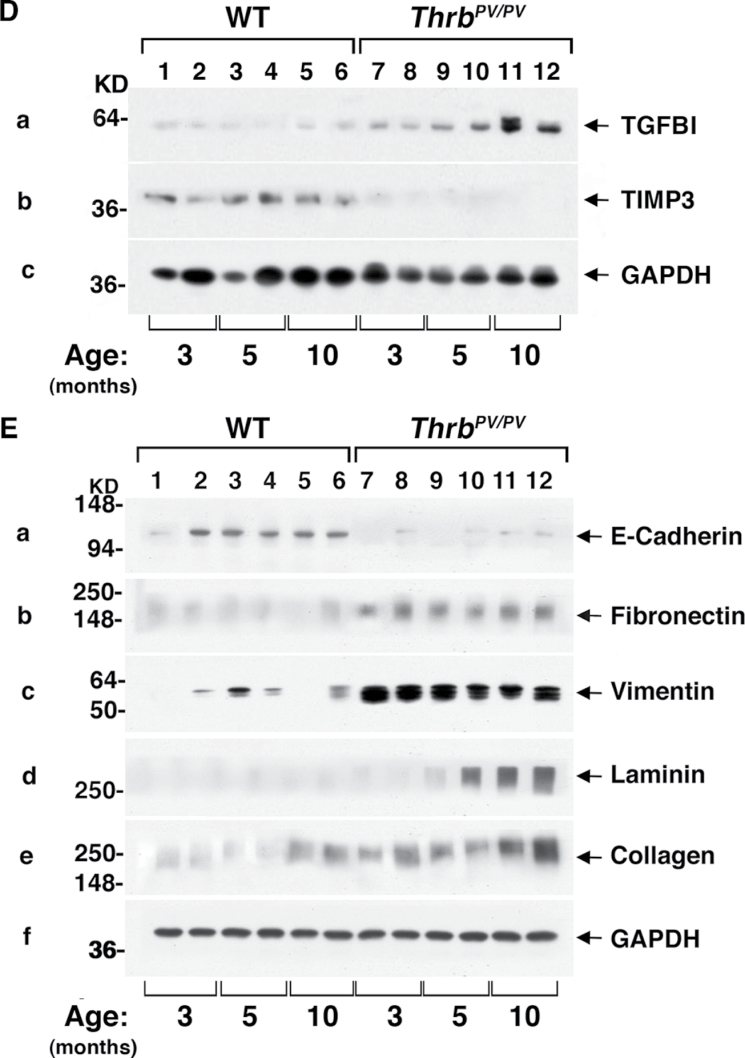
Fig. 3.
Activation of TGFβ-mediated signaling pathway in thyroids of Thrb PV/PV mice. (A) Total tissue extracts of thyroids from WT and Thrb PV/PV mice were prepared as described in Material and methods. Thirty micrograms of total protein was used to determine the protein abundance of TGFBRII (A-a), the phosphorylated form of SMAD2/3 (Ser423/425) (A-b) and total SMAD2/3 (A-c) by western blot analysis. GAPDH was used as a loading control (A-d). (B) Immunohistochemical analysis of TGFβRII and pSMAD2/3 in thyroid tumors of Thrb PV/PV mice. (B-I) Each paraffin section obtained from WT (a, c, e and g) and Thrb PV/PV mice (b, d, f and h) was subjected to immunohistochemical analysis using anti-TGFBRII (panels a and b), anti-pSMAD2/3(Ser423/425) (panels c and d), no primary antibody (negative control; panels e and f) and anti-Ki-67 antibodies (panels g and h). Arrows point to the positive staining for each antibody. (B-II) The positively stained cells were counted, and the data are shown as % of positively stained cells versus total cells (n = 3). The genotypes are marked. (C) Comparison of serum levels of TGFβ1 in Thrb PV/PV and WT mice. The serum concentrations of TGFβ1 were determined, as described in Materials and methods, using sera obtained from WT and Thrb PV/PV mice at the indicated age. Data are expressed as mean ± standard error of the mean (n = 6–13) with P values shown. NS, not significant. (D) Thyroid extracts (30 μg) of WT and Thrb PV/PV mice cells were analyzed by western blot analysis using anti-TGFBI (panel a), anti-TIMP3 (panel b) and anti-GAPDH as a loading control (panel c). (E) Analysis of protein abundance of key regulators in EMT in the thyroid tumors of Thrb PV/PV mice. Thyroid extracts (30 μg) of WT and Thrb PV/PV mice cells were analyzed by western blot analysis using anti-E-cadherin (panel a), anit-FN1 (panel b), anti-vimentin (panel c), anti-Laminin (panel d), anti-collagen (panel e) and anti-GAPDH as a loading control (panel f).


