Fig. 3.
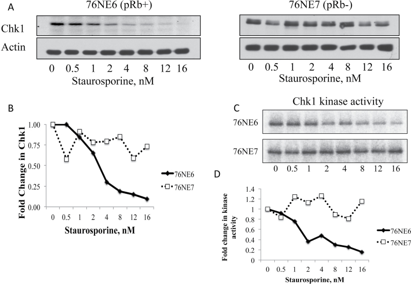
ST inhibits Chk1 in pRb+, but not Rb−, cells. Chk1 expression was measured by (A) western blot in 76NE6 and 76NE7 cells that were treated with increasing concentrations (0–16nM) of ST. (B) Values from densitometric analysis of three independent trials were normalized to the untreated controls and graphed as fold change. (C) Chk1 was immunoprecipitated from the ST-treated 76NE6 and 76NE7 cells and the kinase activity was measured against GST-CDC25a. (D) The densitometry from the average of two independently performed kinase assay blots was graphed.

