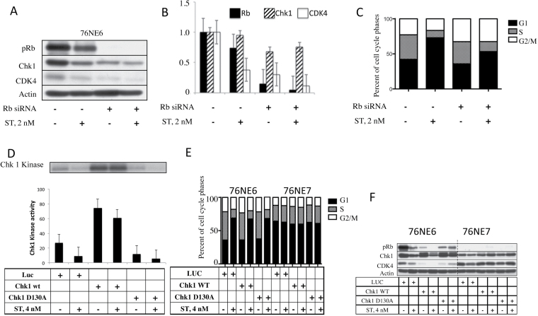
Fig. 4.
ST requires pRb to induce G1 arrest and cannot be reversed when Chk1 is expressed. 76NE6 cells were incubated with 30nM siRNA against Rb for 24h and then treated with 2nM ST and subjected to (A) western blot followed by (B) densitometry. Each experiment was set up twice, and the bar graph shows the average of the two trials. (C) Flow cytometry using PI staining was used to assess the cell cycle distribution of cells. The graph shows the average of three trials. 76NE6 cells were infected with adenoviral LUC or Chk1 and treated with 4nM ST. (D) Cell lysates were immunoprecipitated for Chk1 to measure the kinase activity against GST-Cdc25a. The average of two quantifications by phosphorimaging is shown. (E) Flow cytometry was performed on PI-stained 76NE6 and 76NE7 cells to determine the percentage of cells in each cell cycle phase after infection with either Chk1 or LUC adenovirus and ST treatment. (F) Western blots were used to assess the expression of pRb, Chk1 and CDK4 after Chk1 overexpression and ST treatment.

