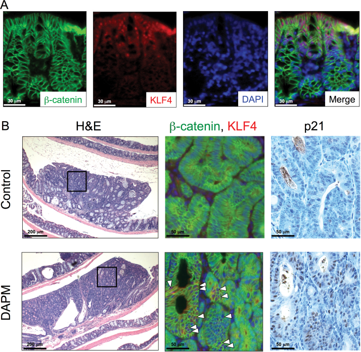
Fig. 5.
β-Catenin, KLF4 and p21 expression in AOM-induced colon tumors. DAPM was administered to A/J mice following AOM treatment as described in Materials and methods. Tissue sections were prepared from the colon of control (n = 15) and DAPM-treated mice (n = 15) and processed for immunofluorescent and immunohistochemical analyses as described in Materials and methods. (A) Double immunofluorescence staining for β-catenin (green) and KLF4 (red) is shown in normal epithelium adjacent to a colon tumor from untreated control mouse. Nuclei were counterstained with DAPI (blue). Merged images represent the overlay of the β-catenin, KLF4 and DAPI staining. (B) Hematoxylin and eosin, β-catenin, KLF4 and p21 staining are shown for tumors from control and DAPM-treated mice. The boxed areas in hematoxylin and eosin sections are enlarged to show areas of positive staining for β-catenin, KLF4 and p21. White arrowheads indicate the KLF4-positive cells within the tumor epithelium. Each serial section was subjected to immunohistochemical analysis of p21.

