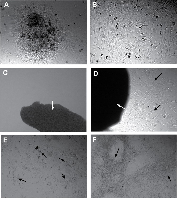
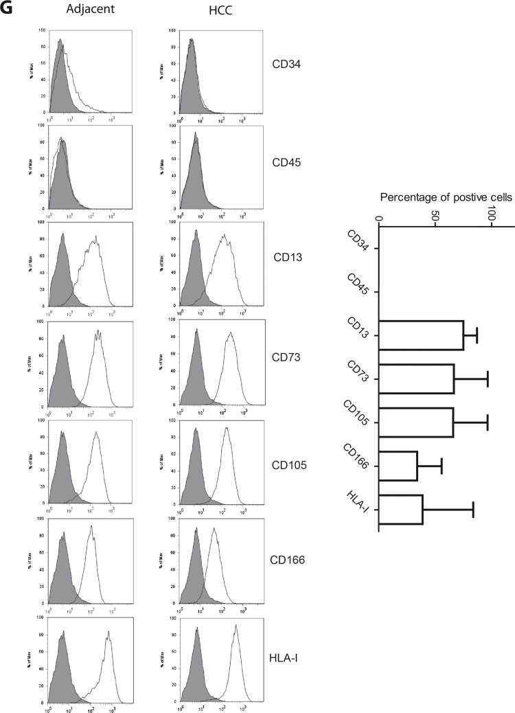
Fig. 1.
Culture and characterization of MSCs from human HCC tissues. After collagenase digestion of surgical resected human HCC tissues and culturing, colonized cell clusters appeared (A) and these cells could rapidly grow and expand by subculture, showing typical fibroblast-like morphology (B). Using another method of culturing tiny tissue specimens, MSC-like cells could not be obtained from adjacent liver tissues (C) but only could be obtained from tumor tissues (D). (E) Adipogenic differentiation of liver carcinoma-derived MSCs, detected by Oil Red O staining for lipid droplets (arrow). (F) Osteogenic differentiation of these cells was evaluated by detection of deposited calcium phosphates using Alizarin Red S staining (arrow). (G) Fluorescence-activated cell sorting and staining confirmed that these cells are positive for the common mesenchymal markers CD13, CD73, CD105 and CD166 and are negative for the common hematopoietic markers CD34 and CD45. The percentages of cells positive for tumor-derived MSCs are presented as mean ± SD (n = 6).


