Figure 4.
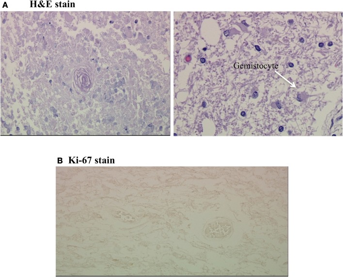
(A,B) Coagulative necrosis and negative Ki-67 stain. (A) Shows two H&E stains from the grossly necrotic region; the first demonstrates coagulative necrosis with a hyalinized fibrotic blood vessel in the center and the second demonstrates scattered gemistocytes on a background of edematous white matter. (B) is a Ki-67 stain of this region, which was completely negative.

