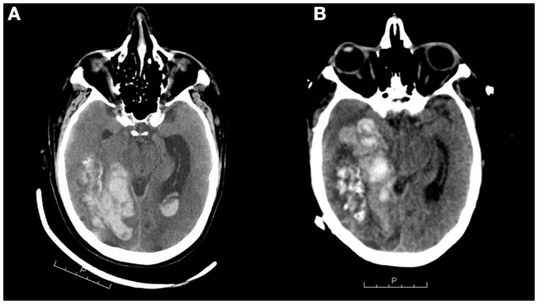
Figure 2.
CT scans (A,B) demonstrating intracranial hemorrhage 3 years after initial SAH. The left scan shows a large right temporal hematoma that was emergently evacuated via craniotomy. The right scan, taken 1 month later, shows extensive intraparenchymal hemorrhage centered in the right temporal lobe extending into the right parietal and occipital lobes measuring up to 7.8 cm × 3.7 cm. There is bleeding into the ventricular system. Note the mass effect, causing midline shift toward the left of 7 mm and uncal herniation.

