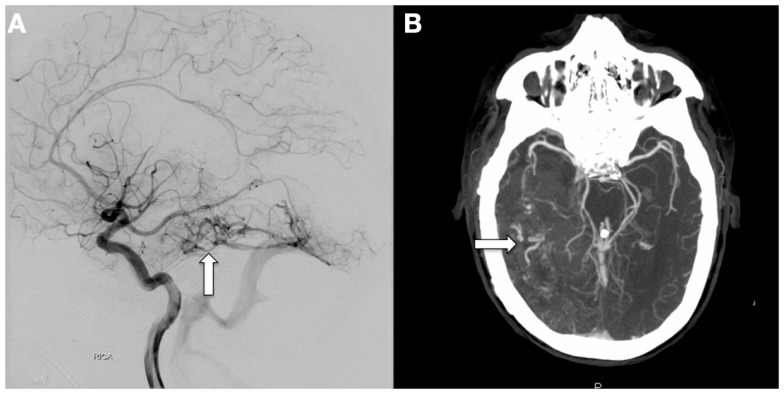
Figure 3.
Catheter-based, diagnostic cerebral angiogram (A) and CT-angiogram (B) taken following the CT scans on the left and right of Figure 2, respectively, showing a large tangle of abnormal vessels centered in the right temporal lobe covering an area of at least 7.8 cm in the AP dimension (arrows). The right P2 segment appears to feed a portion of this vascular malformation, and an enlarged vein appears to drain into the basal vein of Rosenthal. These findings are suggestive of AVM.

