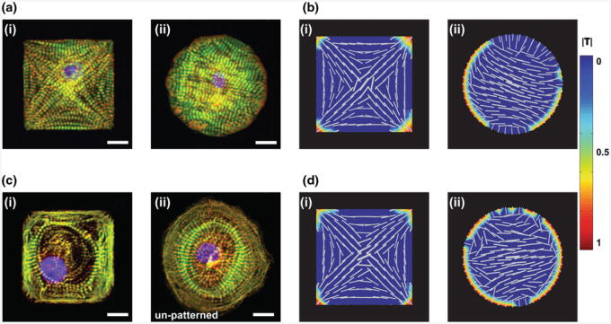
Fig. 3.
Comparison of myofibrillogenesis observed in primary and stem cell-derived cardiomyocytes in vitro with in silico simulations a NMVM on patterned FN at day 3 after seeding (i) square, (ii) circle; b Computational model of a (i) square and (ii) circular cell with myofibril mutual alignment turned on showing polarization in both cell shapes; c human iPS-derived cardiomyocytes on (i) patterned FN square, and (ii) isotropic FN; d Computational model of a (i) square and (ii) circular cell with myofibril mutual alignment turned off showing polarization only in the cell type with non-homogenous boundary conditions; (a, c) scale bar = 10μm; (b, d)color bar shows normalized traction stress (|T|) see Grosberg et al. (2011) for details on the model

