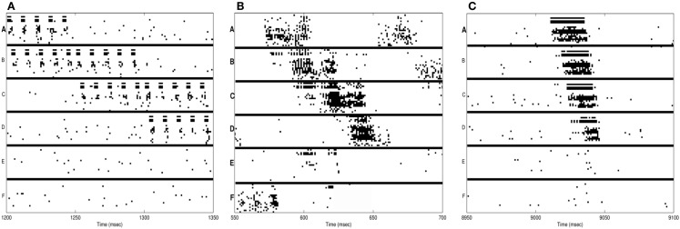
Figure 3.
Neuron activations during procedural learning task. (A) Spike raster plot of the training phase. Stimuli A and B are followed by the stimuli B and C, which are followed by C and D. (B) Spike raster plot showing the activation of the correct sequence during sleep. (C) Spike raster plot showing the activation of the correct sequence after a single session of wake-training followed by renormalization in sleep.

