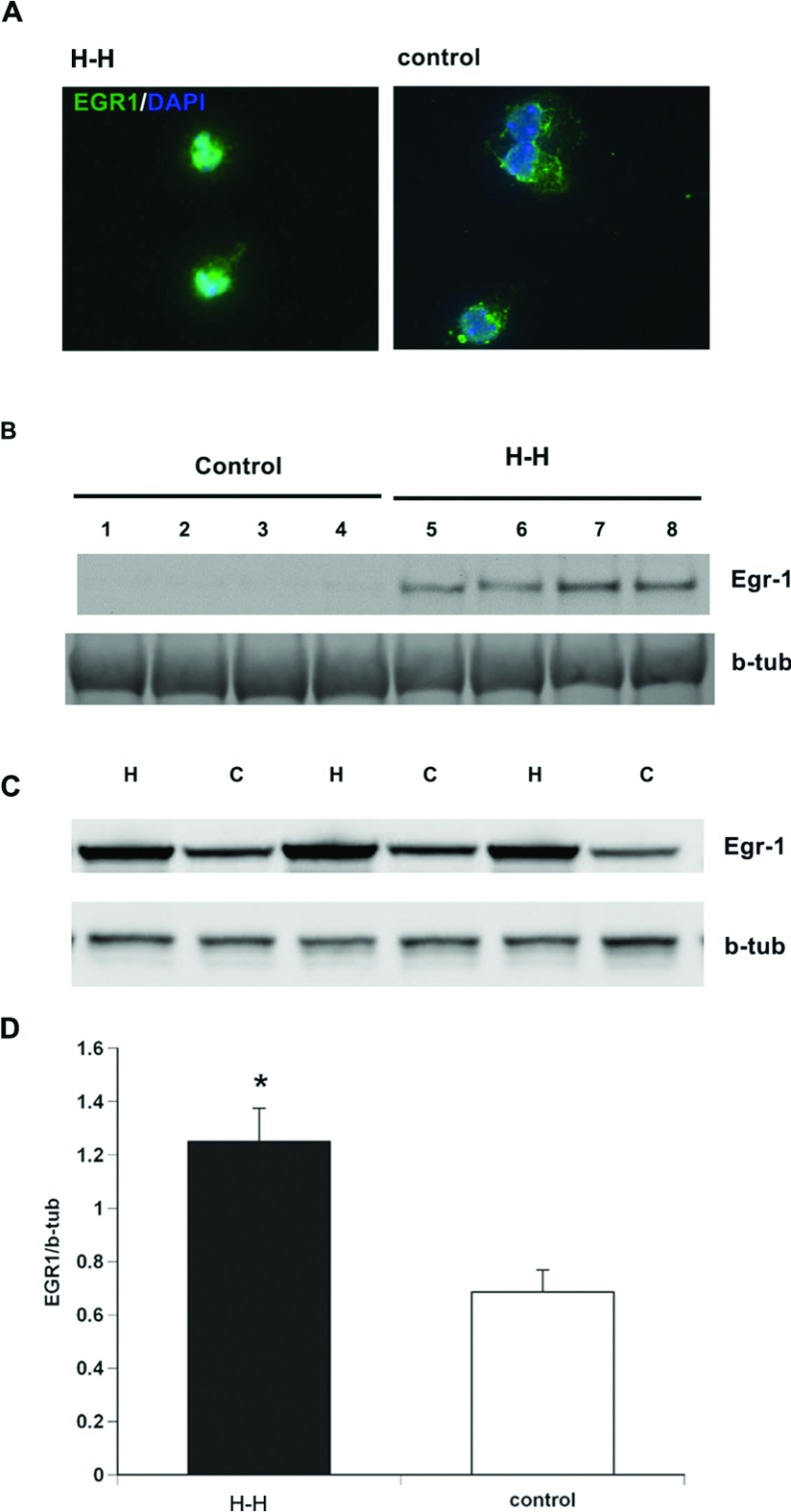
Figure 4. Egr-1 accumulates in the nucleus following H–I and binds the EGF-R promoter.
(A) Rat neurospheres were subjected to H–H and stained for Egr-1 (green) and counterstained with DAPI (blue). (B) Nuclear extracts from neurospheres subjected to H–H and control neurospheres and probed with antibodies specific to Egr-1. (C) Egr-1 levels in NPs subjected to hypoxia alone compared with control NPs. (D) Densitometric quantification of Western blots. Values represent mean±S.E.M.*P<0.05 by Student's t test.

