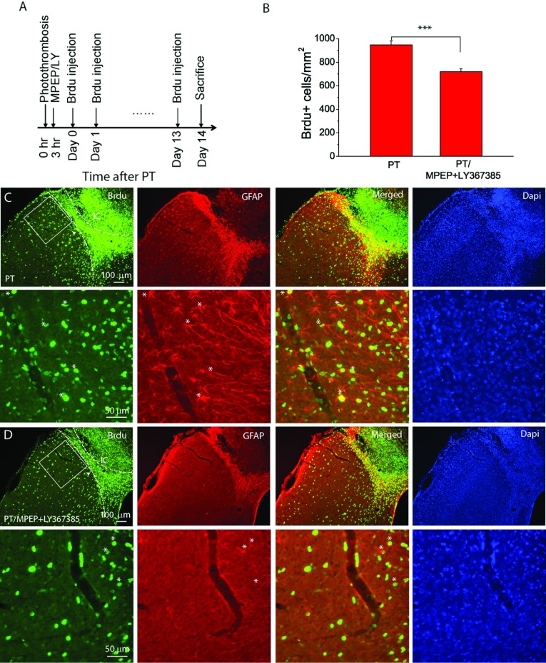
Figure 7. MPEP and LY367385 attenuate cell proliferation in the peri-infarct region after PT.
(A) Experimental design of BrdU injections for cell proliferation study. BrdU+ was injected in mice once a day starting the same day after PT for 14 times and mice were killed 1 day after last injection. (B) Summary of the density of BrdU+ cell in a peri-infarction region of 325 μm×325 μm in the layer 2/3 of cortices (see boxed region in (C,D). The data were averaged from N=7 mice for each group. *P<0.0001, t test. (C,D) Fluorescent images of BrdU and GFAP staining in the infarct and peri-infarct regions of mice not subject (C) or subject (D) to MPEP and LY367385 injection. The left-hand dashed lines indicate boundary of dense BrdU+ cell, and the right-hand dashed lines indicate the boundary of proliferating cells in glial scars based on GFAP staining. IC, ischaemic core. The bottom panels in (C) and (D) are the high-resolution images of the boxed regions in the upper panels and some of the BrdU+/GFAP+ cells are indicated by *. Note there are more BrdU+/GFAP+ cells in (C) than in (D).

