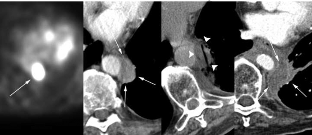
Figure 2.
The same 68-year-old patient was found to have an FDG PET-positive left para-aortic node (first image) 4 months after the ablation procedure described in Figure 1. This enhancing lesion measuring 1.8 × 3.5 × 4.2 cm (second image) underwent cryoablation (third image), resulting in a resorbed hypodense non-enhancing soft tissue site on follow-up axial CT (fourth image). As demonstrated in the third image, well-demarcated hypodense margins of the iceball allow safe ablation adjacent to crucial structures such as the aorta.

