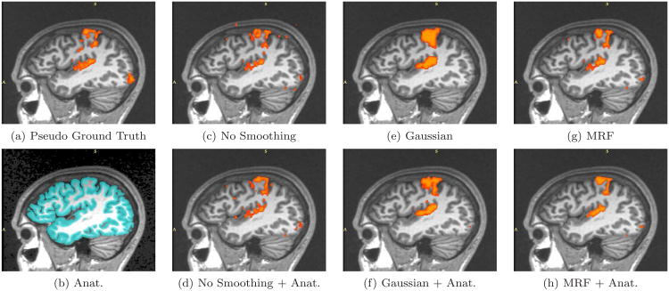
Fig. 9.
Real fMRI study. One sagittal slice in the estimated activation maps. (a) Pseudo ground truth created from four full-length sessions (17 epochs each). (b) High resolution segmentation result overlaid onto the corresponding MR image. (c)-(h) The activation maps obtained from the first seven epochs. (c) Plain GLM detection results. (d) A gray matter mask is employed to suppress GLM-detected activations which do not lie in the gray matter. (e) Isotropic Gaussian smoothing with 7mm FWHM is applied prior to the GLM detector. (f) A weighted Gaussian smoothing is applied prior to the GLM detector. (g) is generated by the GLM detector with MRF spatial prior. (h) further incorporates the anatomical information into the MRF prior.

