
Figure 3.
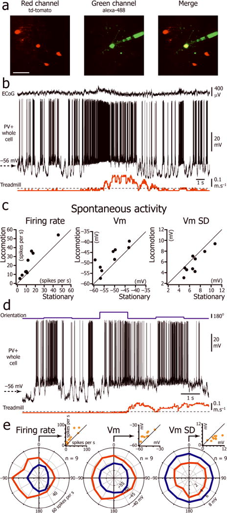
Effect of locomotion on Vm of L2/3 parvalbumin positive interneurons. (a) In vivo two-photon image of a neuron (yellow) injected with Alexa-488 (green) during the recording in a mouse expressing tdTomato (red) in PV+ neurons. Scale bar: 50 μm. (b) Spontaneous activity of a L2/3 PV+ interneuron during immobility and locomotion. (c) Plots of firing rate, Vm and Vm SD during immobility versus locomotion for 9 L2/3 PV+ interneurons (8 mice). (d) Vm of the neuron shown in c during the presentation of drifting gratings of three different orientations (top trace) interleaved with presentation of an isoluminant gray screen. (e) Orientation tuning curve of the L2/3 PV+ interneuron population for firing rate, Vm, and Vm SD during immobility and locomotion (n=9 neurons from 8 mice). The orientation “0” was assigned for each neuron to the orientation at which the stationary firing rate evoked by the visual stimulus was maximal. Insert: Plot of firing rate, Vm, and Vm SD during immobility versus locomotion for the orientation “0”.