Figure 1.
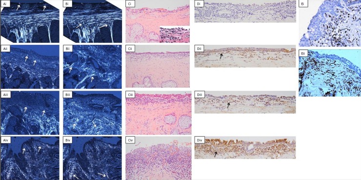
Example images for conjunctival histological grading. (A) Connective tissue scarring in the subepithelial space. Cross-polarised light is used. (i) Shows normal tissue (grade 0) with collagen fibres parallel to the surface (arrows). (ii–iv) Show progressive disorganisation of this appearance (grades 1–3) with the arrows indicating the subepithelial collagen fibres. Original magnification ×100. (B) Connective tissue scarring in the tarsal tissue. Cross-polarised light is again used. (Bi) shows normal tissue (grade 0) with long collagen fibres between the meibomian glands which join shorter fibres next to the stroma forming a ‘T’ sign in normal tissue (arrows). (ii–iv) Show progressive disorganisation of this appearance (grades 1–3). Original magnification ×100. (C) Inflammatory cell infiltrate in the subepithelium. Heamtoxylin and eosin stain used, arrows show inflammatory cells. (i–iv) Correspond to grades 0–3. The inset in (Ci) shows the inflammatory cells at a higher magnification. Original magnification ×200. (D) CD83 cell in the subepithelium. These are stained brown as shown by the arrows. (i–iv) Correspond to grades 0–3. Original magnification ×200. (E) CD45 cells with a dendritic morphology. These are also stained brown but have a dendritic morphology, as shown by the arrows. Original magnification ×400.

